Kick-off for the DISAIP research project: AI for improved stroke diagnosis with reduced radiation exposure
In the event of a stroke, every minute counts. Doctors need to identify quickly which areas of the brain can still be saved. To do this, they use perfusion CT: a contrast agent is injected, and the brain is scanned repeatedly over a short period to reveal how well the individual areas are supplied with blood. However, the large number of scans results in high radiation exposure. A lower dose would introduce noise into the images and make diagnosis more difficult. Previous methods for improving image quality have relied solely on the perfusion scans themselves.
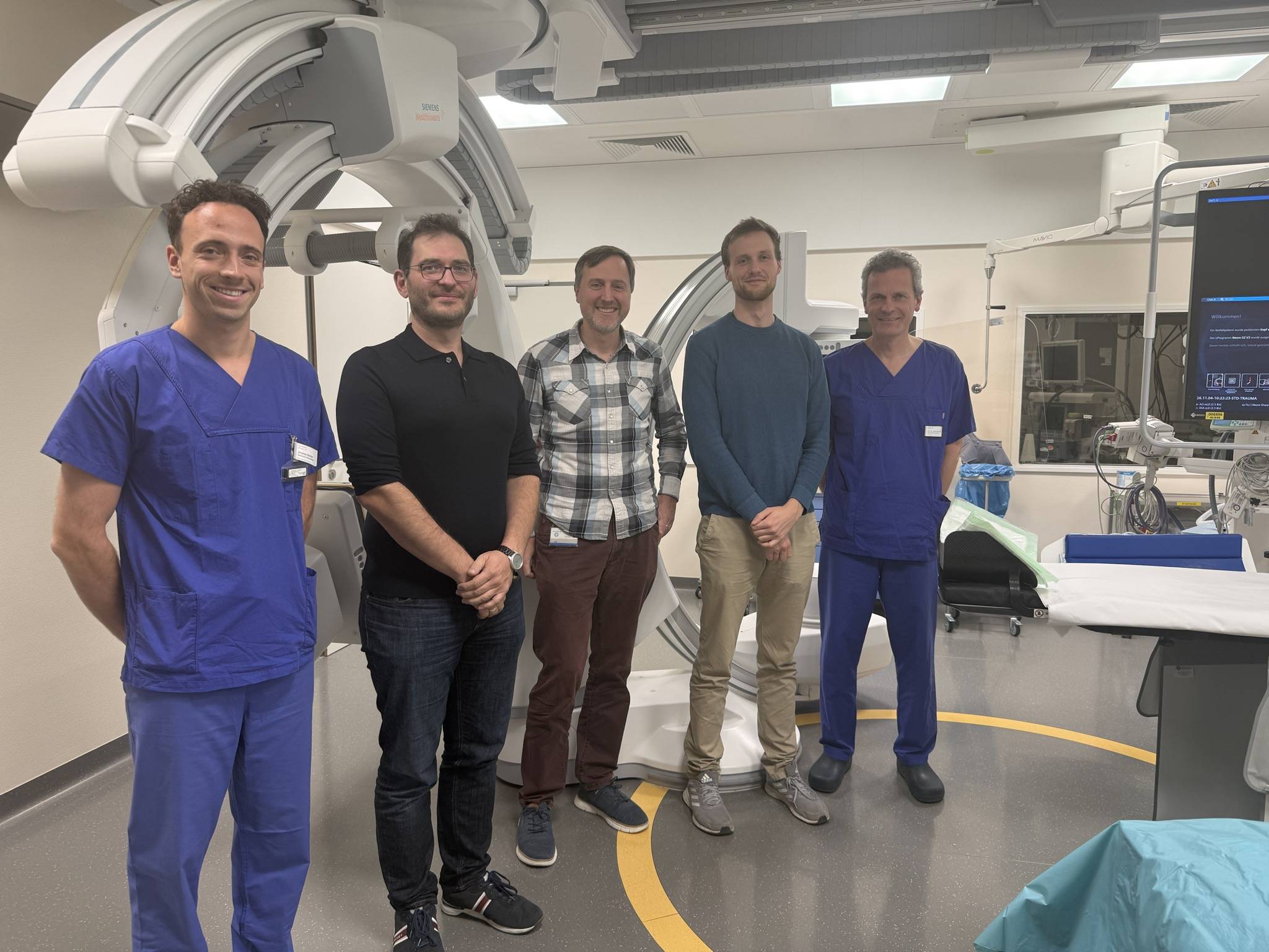
This is where the new research project DISAIP (Dose Reduction and Improved Stroke Diagnosis with Multi-Source AI Denoising of Perfusion CT) comes in. It is being carried out jointly at Günzburg District Hospital, the University of Ulm and the Technical University of Ulm (THU), and was launched with a kick-off event on 4 November. Funded by the German Research Foundation (DFG) as part of a DFG grant, the project is pursuing a new approach: artificial intelligence is to additionally incorporate high-resolution CT images, which are acquired as standard in stroke patients anyway. By combining both image sources, it aims to deliver sharper, lower-noise perfusion images that reveal finer details of cerebral blood flow. At the same time, this allows the radiation dose to be reduced. The aim is to achieve a more accurate diagnosis with lower radiation exposure.
The THU team, led by Prof. Dr Alfred Franz, is developing 3D-printed perfusion phantoms as part of the project. These are artificial replicas based on real patient anatomies, which allow blood flow to be simulated. They enable the AI to be tested under realistic conditions and using known reference values, without placing any additional burden on patients.
“With the 3D-printed perfusion phantoms, we are creating a realistic yet controllable basis for testing the AI under defined conditions. In this way, we can help to verify the performance of the new imaging method and, in the long term, improve stroke diagnostics,” explains THU Professor Dr Alfred Franz.